
For millions of patients around the world, routine medical procedures such as intravenous line insertions, blood draws, and medication delivery evoke significant stress and physical discomfort. The challenge often stems not from the needle itself, but from the difficulty healthcare providers face in locating a suitable vein beneath the skin. Factors such as dehydration, age, repetitive treatments, obesity, or vascular disease can render peripheral veins fragile, rolling, or virtually invisible to the naked eye. In response to these clinical hurdles, near-infrared vascular imaging technology—most notably embodied by devices such as the VeinViewer—has reshaped the standard of vascular access, transforming an invasive guessing game into an exact visual science.
“Sci-Fi Made Reality” Highlights
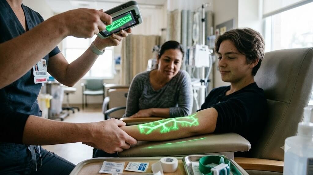
- Augmented Reality on Human Skin: Like a medical heads-up display (HUD) from a cyberpunk film, it uses real-time digital projection to turn the patient’s arm into a live, illuminated anatomical screen.
- Invisible Light, Visible Results: Utilizes invisible near-infrared light spectrums (invisible to the human eye) that interact with internal biochemistry and bounce back into visible high-contrast wavelengths.
- X-Ray Vision Without Radiation: Gives clinicians the functional equivalent of sub-dermal “X-ray vision” to map venous pathways up to millimeters below the surface without any harmful radiation or physical contact.
- Predictive Navigation: Allows doctors and nurses to spot anatomical obstacles—such as deep bifurcations, rolling vessels, or hidden valves—before ever unboxing a needle.
Core Scientific & Technical Highlights
- Biophysical Mechanism: Near-infrared light is selectively absorbed by deoxygenated hemoglobin in venous blood while being scattered and reflected by surrounding fatty and cutaneous tissue.
- Real-Time Projection Mapping: High-speed sensors capture the optical differential, an onboard digital processor maps the vessel coordinates, and an aligned visible-spectrum projector casts the exact vascular layout onto the skin in real time.
- Clinical Accuracy: Detects peripheral veins, assesses patency, visualizes real-time blood flow, and catches vessel infiltration or hematomas immediately.
- Patient Outcome Metrics: Significantly increases first-stick cannulation rates, halves procedural search time, and dramatically cuts procedural pain, bruising, and phobia in pediatric, oncology, and geriatric care.
The Biophysics of Near-Infrared Vascular Mapping
At the core of the VeinViewer system lies an elegant application of optical physics and real-time digital projection. The device does not rely on invasive radiation or ultrasound gel; rather, it harnesses harmless near-infrared (NIR) light emitted directly onto the patient’s skin.
Human tissue and blood interact with NIR light in distinct manners:
- Hemoglobin Absorption: The deoxygenated hemoglobin circulating within venous blood absorbs near-infrared wavelengths efficiently.
- Tissue Reflection: The surrounding connective tissue, subcutaneous fat, and epidermal layers do not absorb this wavelength to the same degree, instead scattering and reflecting the light back toward the device.
Sensors embedded within the scanner detect these differences in optical reflection and feed the data into an onboard digital processing unit. The processor rapidly computes an accurate map of the vascular architecture beneath the surface. It then projects this processed image back onto the skin in real time using visible light (often green or high-contrast illumination). Because the projection aligns precisely with the patient’s anatomy, clinicians observe a detailed, glowing roadmap of veins appearing directly on the skin’s surface.
Clinical Precision: Beyond Surface Visibility
Traditional vein identification relies primarily on palpation—feeling the bounce and resilience of a vein through the skin. While experienced clinicians develop strong tactile instincts, palpation cannot reveal underlying anatomical anomalies or internal blood flow dynamics.
Advanced near-infrared imaging reveals critical parameters that touch alone cannot assess:
- Depth and Diameter: Clinicians can evaluate vein caliber and trace vessels up to several millimeters beneath the surface, ensuring the chosen vein is large enough to accommodate the required catheter gauge.
- Vascular Course and Bifurcations: The visual projection reveals curves, branching points, and junctions where valves might otherwise obstruct a catheter’s advancement.
- Vessel Patency and Hematomas: Real-time feedback allows clinicians to observe vessel collapse, hematoma formation, or sluggish flow, prompting them to avoid compromised sites before attempting an insertion.
By providing dynamic visualization before, during, and after venipuncture, clinicians can verify successful vessel cannulation and detect immediate infiltration or extravasation, avoiding prolonged tissue damage.
Mitigating Patient Trauma and Procedural Anxiety
The clinical benefits of vascular illumination translate directly into improved patient experience. Repeated attempts to establish an IV line—known as “difficult venous access” (DVA)—frequently lead to severe bruising, phlebitis, nerve irritation, and lasting needle phobia.
In specialized demographics, the impact is particularly pronounced:
- Pediatric Care: Children often experience intense fear during needle-based procedures. Shortening procedure time and achieving first-stick success dramatically lowers stress for both the child and their parents.
- Geriatric and Oncology Patients: Fragile, sclerosed veins resulting from advanced age or repeated chemotherapy regimens make traditional access difficult. Visual guidance preserves viable vessels and prevents unnecessary trauma.
- Emergency and Critical Settings: In high-acuity environments where rapid fluid resuscitation or medication administration is vital, eliminating delays caused by failed access attempts can be lifesaving.
Studies consistently demonstrate that implementing optical vein-finding systems increases first-attempt success rates, reduces the number of needle sticks per patient, and cuts procedural time in half.
Operational Efficiency and Health System Impact
Beyond bedside comfort, vascular visualization delivers measurable institutional advantages. Repeated cannulation attempts consume substantial supplies, including needles, dressings, extension sets, and local anesthetics. Furthermore, prolonged attempts divert nursing and physician hours away from other critical tasks.
By standardizing access procedures, hospitals experience:
- Decreased catheter-related complications and infections.
- Lower equipment waste associated with discarded cannulas and kits.
- Reduced reliance on central venous catheters or peripherally inserted central catheters (PICCs), which carry higher procedural risks and substantial management costs.
- Higher patient satisfaction scores, which play a central role in hospital quality metrics and clinical accreditation.
The Evolution of Non-Invasive Clinical Diagnostics
The transition from blind or tactile access to projection-based digital assistance reflects a broader paradigm shift in modern medicine: reducing invasiveness while increasing diagnostic clarity at the point of care. As optical imaging, machine learning, and miniaturized projection hardware advance, future iterations of vascular visualization will offer even greater depth penetration, automated vein selection algorithms, and integration into handheld consumer and point-of-care ultrasound devices.
By bridging optical physics and daily clinical practice, technologies like VeinViewer remove uncertainty from vascular access, replacing a source of routine distress with a safe, precise, and gentle standard of care.
Frequently Asked Questions (FAQ)
What is VeinViewer, and how does it work?
VeinViewer is a non-invasive imaging device that uses near-infrared (NIR) light to create a real-time visual map of a patient’s vasculature directly on their skin. The deoxygenated hemoglobin in blood absorbs the infrared light, while the surrounding tissue reflects it. The device captures this contrast, processes it digitally, and projects a visible map showing precisely where viable veins are located.
Does VeinViewer touch the patient or emit harmful radiation?
No. VeinViewer is completely non-invasive and operates from a short distance above the skin. It does not use ionizing radiation or require conductive gels, making it safe and clean for repeated clinical use across all patient populations.
How does vascular illumination reduce needle pain?
The pain associated with blood draws and IV placement often escalates when veins are difficult to find, leading to multiple failed attempts or vessel damage. By providing a clear roadmap of vessel depth, width, and direction, the technology allows healthcare providers to achieve successful cannulation on the first stick, significantly minimizing physical discomfort and anxiety.
Which patients benefit most from vein-finding technology?
While beneficial for all patients, it is especially valuable for individuals with difficult venous access (DVA). This includes pediatric patients, elderly individuals with fragile veins, oncology patients undergoing chemotherapy, individuals with deeper veins or obesity, and patients suffering from severe dehydration.
Can VeinViewer prevent complications like hematomas or infiltration?
Yes. Beyond locating veins for initial puncture, the live projection allows clinicians to monitor vessel patency and fluid flow in real time. If a vein collapses, forms a hematoma, or leaks medication into surrounding tissue (extravasation), the practitioner can detect it immediately and take corrective action before complications worsen.